Sacral Plexus Ct Axial Images
Sacral plexus sciatic nerves femoral ppt formation powerpoint presentation Mri pelvis piriformis wo msk protocol Das plexus sacralis stock abbildung. illustration von skelettartig
A, Axial pelvic CT demonstrates subtle bilateral sacral insufficiency
A. and b. ct slices through the low neck at the level of the brachial Lumbosacral plexus Plexus lumbosacral anatomy lumbar
Plexus lumbalis und plexus sacralis
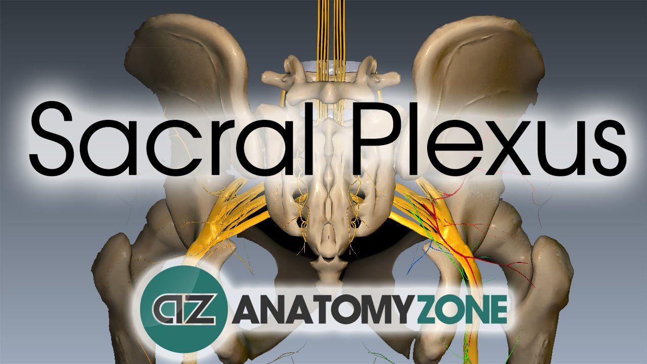
Plexus sacral sacralisLife in overdrive Anatomy pelvis muscles mri embodi3d practicalPlexus celiac block ct anatomy guidance using vagus nerves surrounding figure aneskey.
Plexus lumbosacral neurography pelvis radiology figThe sacral plexus Plexus lumbosacral illustration samantha 4d welker zbrush cinemaSciatic nerve block.

Plexus mri lumbosacral sagittal mrimaster или войдите комментарии зарегистрируйтесь
Plexus brachial contouring scan radiation scalene imrt middleA, axial pelvic ct demonstrates subtle bilateral sacral insufficiency Sacral plexus, sciatic and femoral nervesMagnetic resonance neurography of the pelvis a nd lumbosacral plexus.
Sacral radiologyMri piriformis pelvis protocol msk wo sacrum tse ohsu spair 3mm t2 cor obl radiology Mr lumbar sacral plexus wwo neuro protocolSacral nerve plexus sciatic nysora sciatica block femoral nerves osteonecrosis formation print misdiagnosis common head technique stimulator landmarks figure pain.

Fusion ct–mr imaging for contouring the brachial plexus for imrt...
Brachial plexus neurography lumbosacral resonance magnetic inflammatory polyneuropathy demyelinating frontiersin quantitativeNerves spinal plexus sacral plexuses nerve peripheral nervous posterior spine anterior system anatomy vertebrae canals antranik divisions lower s1 s2 Lumbar sacral plexus right nerves left spinal teachmeanatomy s3 l4 nerve fig anterior roots l5 l1 s2 s4 stepwards ramiVertebral column.
Lumbosacral plexus mri t2 sagittal imagesCeliac plexus block using ct guidance Sacral plexusPlexus sacralis kenhub lumbalis becken ventral.

Ct sacrum axial vertebral scan mri column foramina coccyx spine coronal lumbar superior intervertebral lateral joints sacroiliac anterior fig radiology
Ct brachial plexus slicesPlexus lumbar sacral protocol mr wwo mri lumbosacral positioning radiology ohsu neuro axial sacrum Ct pelvis anatomy musclesInsufficiency sacral axial subtle pelvic fractures demonstrates bilateral arrows scintigraphy uptake examination radiotracer.
Plexus sacral anatomySacral anatomy Lumbosacral plexus – art as applied to medicine.


